Acoustic Neuroma

Introduction

Following cell differentiation and proliferation, cell division is the process through which cells are multiplied. Tissues are groups of cells, and the abnormal growth of tissue in an organism is referred to as a tumour. Tumours typically originate as a result of certain disruptions in cell development and the generation of new cells. When a tumour’s growth is restricted, it is benign (non-cancerous), but when it spreads to the body’s key organs, it is malignant (cancerous).

What is Acoustic Neuroma?

Acoustic neuroma is a non-malignant and rare tumour that is also called a Vestibular schwannoma. It is produced by the Schwann cells that surround and support the nerves. The vestibular and auditory nerves, which control balance and hearing, respectively, compose the branches of cranial nerve VIII, commonly known as the vestibulocochlear nerve, where tumours have grown. A critical instance develops when the tumour grows rapidly and continuously.

Causes of Acoustic Neuromas

  • Some people have a rare genetic condition called neurofibromatosis type 2, which is characterized by the formation of tumours on the nerves. Acoustic neuroma is a result of this condition. 
  • Acoustic neuromas are reported in only 5% of patients with neurofibromatosis type 2 (NF2 patients). 
  • In the majority of cases, the exact aetiology of auditory neuroma is unknown. However, some risk variables, including family history, radiation exposure, age, and loud noise exposure, are still thought to be the root cause.

Symptoms of Acoustic Neuroma

Along with other difficulties, the growth of tumours in the vestibulocochlear nerve might affect balance. The following are the symptoms of such tumorous growth:

  • Impaired hearing: Acoustic neuromas 90% of the time accompany some degree of hearing loss. The tumour’s pressure on the nerve or the discharge of compounds harmful to hearing can both cause hearing loss. 
  • Tinnitus: Patients with tinnitus experience a high-pitched hissing or buzzing sound in their ears. Tinnitus can occasionally become persistent. Hearing loss may or may not be present in tinnitus patients.
  • Vertigo and loss of balance: Vertigo, a sudden sensation of the head tilting and spinning, is caused by the growth of a tumour on the balance and auditory nerve. Because of this patient can become unsteady and lurch. 
  • The fullness of the ear: Acoustic neuroma patients may experience full ears as if water is trapped in the ear canal. Hearing loss is frequently to blame for this.
  • Other signs and symptoms of an acoustic neuroma include facial numbness, headaches, nausea, changes in taste, and difficulty swallowing.

Diagnosis of Acoustic Neuroma

The examination of the ear is typically the first step in the diagnosis of an acoustic neuroma, which is then followed by evaluations of the patient’s medical history, imaging, and hearing capacity. Tumours in the brain may be detected with MRI or CT scans using magnetic resonance imaging (MRI) or computerized tomography (CT). The following tests are crucial for determining the presence of an acoustic neuroma: 

  • Audiometry: An audiometer uses a painless hearing test to quantify one’s hearing depending on how loud sounds are and how quickly they vibrate. 
  • Pure Tone Average (PTA): it is a measurement used to assess hearing impairment for speech comprehension. A higher rating denotes a hearing impairment.
  • Speech Reception Threshold (SPT):  The patient can hear speech at this volume at least 50% of the time. A higher score, similar to PTA, denotes hearing impairment.
  • Discrimination in speech (SD): It is a test of the patient’s capacity to distinguish between speech in quiet and noisy settings. Hearing loss is indicated by the lower score.

Treatment for Acoustic Neuroma

The course of treatment for an acoustic neuroma might vary; it is typically determined by the patient’s general health, the size and progression of the tumour, and its symptoms. Three treatment methods are available:

  • Monitoring: Adults who have primary slow-growing tumours may not exhibit any symptoms, making patient surveillance a valuable alternative for follow-up care. The ideal situation for a monitor is when the tumours are up to 1.5 cm in size. Before the tumour grows to a dangerous size, surgery must be performed to remove it.
  • Surgery: Acoustic neuromas can potentially be treated surgically. The surgical procedure’s main goals are to eliminate the tumour and avoid facial paralysis. Complete excision, however, may not always be possible due to the tumour’s proximity to vital brain regions. This procedure carries the potential for several side effects, including hearing loss, tinnitus, cerebrospinal fluid leakage via the nasal route, face numbness, etc.
  • Radiation therapy: It is a non-surgical option; stereotactic radiosurgery, which is most frequently used, can stop the growth of the tumour and lessen the death of neighbouring cells. With this technique, the gamma rays are directed precisely to the tumour without damaging nearby cells. For patients with big tumours, this treatment is not advised.

Summary

Tumours typically originate as a result of certain disruptions in cell development and the generation of new cells. Acoustic neuroma is a non-malignant and rare tumour that is also called a schwannoma. Acoustic neuromas are reported in only 5% of patients with neurofibromatosis type 2 (NF2 patients). Along with other difficulties, the growth of tumours in the vestibulocochlear nerve might affect balance. Tumours in the brain may be detected with MRI or CT scans using magnetic resonance imaging (MRI) or computerized tomography (CT).

 Frequently Asked Questions

1. How does Stereotactic Radiosurgery Work?
Ans. With the help of a 3D coordinate system, stereotactic surgery may find small targets inside the body and carry out a variety of minimally invasive surgical procedures on them, including biopsy, ablation, lesion, stimulation, injection, implantation, and radiosurgery, etc.

2. Define Audiometry?
Ans. A diagnostic hearing test is called audiometry. The loudness of the tone and the speed of the sound determine one’s capacity to hear it. For the detection of hearing impairment, it is crucial.

3. What is the Speech Reception Threshold?
Ans. The speech reception threshold is the lowest degree of speech hearing at which a person can recognize 50% of spoken words. Each ear has reached its speech reception threshold. It serves as a reference point for supra-threshold tests and serves to validate the thresholds discovered using PTA.

4. What is Tinnitus?
Ans. Patients with tinnitus experience a high-pitched hissing or buzzing sound in their ears. Tinnitus can occasionally become persistent. Hearing loss may or may not be present in tinnitus patients.

A Guide To Composition And Function Of Lymph

Introduction

The human body is a complex structure made up of several organs that are arranged into different systems and are designed to perform specific functions during various life events. To stay healthy, each of these systems needs a constant supply of oxygen and nutrients. The metabolic wastes produced by each of these systems must also be transported to the excretory organs to get expelled from the body. The body’s fluids, which are constantly circulated throughout the body, carry out all of this transportation. These fluids are lymph and blood. This article will go into further detail about lymph.

What is Lymph?

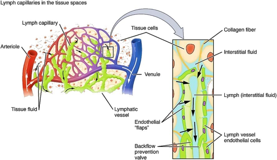
An alkaline translucent fluid called lymph fills the gaps between the lymphatic tissue and travels via the lymph vessels. It is a vascular fluid connective tissue that aids the immune system in performing its duties by transporting chemicals throughout the body. It is the primary component of the lymphatic system.

 Image source

Composition of Lymph

Plasma and lymphocytes are the two components that make up the lymph.

Plasma: The lymph’s fluid component and its matrix are known as plasma. It is produced as a result of the ultrafiltration of blood plasma through the capillary walls. Its composition is essentially similar to that of blood plasma, with the exception that it has a higher concentration of glucose and a lower proportion of proteins, calcium, or phosphorus.

Leucocytes: Unlike blood, the lymph’s cellular makeup only consists of amoeboid WBCs, also known as leucocytes. Lymphocytes are the main form of WBC that are present in the lymph. These WBCs are agranular, and their main job is to help the body’s defence system by assisting in the production of antibodies. So we can say that lymph is the blood that has been stripped of its red blood cells, platelets, and some proteins.

Lymph vs Blood

The two fluid connective tissues in the body are blood and lymph. However, there is a big difference in the way they are made and how they circulate. The distinctions between lymph and blood are outlined in the table below.

LymphBlood
Lymph is a colourless fluid in connective tissue. Blood is a red-coloured fluid. 
The lymphatic system carries lymph through it. The cardiovascular system carries blood through it. 
Leucocytes and plasma make up the lymph. Plasma, leucocytes, erythrocytes, and thrombocytes make up blood. 
Proteins, calcium, and phosphate are present in lymphatic plasma in comparably lower amounts. Higher amounts of calcium, phosphorus, and proteins are present in the blood plasma.

Lymph, Lymphatic Vessels, and Lymphoid Organs

  • The lymphatic system is made up of lymph, lymphatic veins, and lymphoid organs. The interstitial fluid that is lost from the circulatory system as a result of the filtration of blood through capillary walls is restored to it via this supplementary pathway. 
  • The lymphatic system is made up of several soft, fragile lymph nodes, which are bean- or sphere-shaped structures. 
  • Lymph nodes are typically invisible and are hard to find. They can be seen in the armpit, groin, neck, and region between the chest and abdomen. These nodes may swell as a result of an infection within the body, which may be a sign that the body is fighting against the illness.
  • In the lymphatic system, the lymph passes through the lymph vessels. The main lymphatic vessel is the thoracic duct, which enters near the base of the spine and receives lymph from the lower chest, pelvis, and abdomen. 
  • Primary lymphoid organs and secondary lymphoid organs are the two categories of lymphoid organs. The bone marrow and thymus are prominent examples, but the spleen, tonsils, and several other mucus membrane tissues are secondary examples. 
  • The lymphoid organs are linked to the development of immune cells and play a crucial part in the body’s immunological response.

The Function of the Lymph

The lymph has two main roles: supporting the circulatory system and boosting the immune system. The lymphatic system serves the following functions.

  • Immune response: White blood cells, which are a vital component of the body’s defense system, are found in the lymph. Lymphocytes in the lymph secrete antibodies that defend the body against foreign invaders.
  • Fat absorption from the digestive tract: Blood cannot carry fats. They circulate throughout the body after being absorbed by the lymph.
  • Acts as a middle man: Lymph serves as a conduit for the exchange of substances between the blood and the tissues. so serves as a bridge between the blood and tissue cells.
  • Waste material transportation: Waste products leave the tissues and are carried by lymph, which then excretes them into the blood for them to be transported to the excretory organs for removal from the body.

Summary

An alkaline, translucent fluid called lymph fills the gaps between the lymphatic tissue and travels via the lymph vessels. Plasma and lymphocytes are the two components that make up the lymph. The lymphatic system is made up of lymph, lymphatic veins, and lymphoid organs. The lymph has two main roles: supporting the circulatory system and boosting the immune system.

Frequently Asked Questions  

1.Why is Lymph Regarded as Blood without Some Proteins, Red Blood Cells, or Platelets?
Ans. White blood cells and plasma make up the lymph. Although the composition of lymphatic plasma is essentially identical to that of blood plasma, it lacks certain proteins, and coagulating agents, and has significantly less calcium and phosphorus. Lymph can therefore be compared to blood, which lacks some proteins, red blood cells, and platelets.

2.How does Swelling of Lymph Nodes Occur?
Ans. Any type of infection in the body can cause swelling in the lymph nodes. It is a sign that the body’s immune system is working to fight off an infection of some kind.

3.How does the Lymphatic System Function?
Ans. By supporting the immune system and guarding the body against external intruders, the lymphatic system serves a crucial role in the body. Additionally, it controls blood volume by replenishing the interstitial fluid that drains from blood as it flows through blood capillaries.

4.Define Lymphedema.
Ans. Lymphedema is a condition in which the body’s lymphatic system accumulates protein-rich fluid, which causes swelling in tissues. Typically, it affects the hands or legs, but it may also develop on the chest walls, abdomens, necks, and genital areas.

5.What is Lymphatic Cancer?
Ans. Lymphoma is s cancer that affects the body’s immune system, which fights germs. There are lymph nodes (lymph glands), spleens, thymus glands, and bone marrow in the lymphatic system. All of these areas, as well as other body organs, can be affected by lymphoma.