Muscular system

Introduction

The muscular system is an organ system in the body that is responsible for generating force and movement. It includes all the muscles in the body, as well as their associated tendons, which connect muscles to bones, and ligaments, which connect bones to other bones. The muscular system works together with other systems in the body, such as the nervous system and skeletal system, to coordinate movement and maintain posture. The muscular system is important for a variety of bodily functions, including movement, posture, and heat generation. 

Types of Muscular system

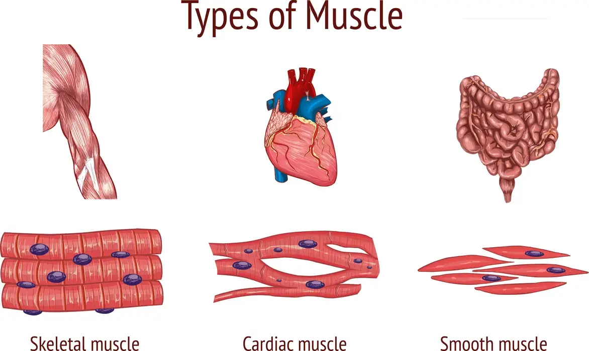
There are three types of muscles in the human body:

  1. Skeletal muscles
  2. Smooth muscles
  3. Cardiac muscles

Diagram showing the types of muscle in the muscular system

The skeletal muscle system

The skeletal muscle system is responsible for generating movement and providing support for the body. Skeletal muscles are attached to bones by tendons and are under voluntary control, meaning that they can be consciously controlled to move. Here are some key features of the skeletal muscle system:

  1. Structure: Skeletal muscles are composed of bundles of muscle fibers that are surrounded by connective tissue. The muscle fibers are made up of myofibrils, which contain actin and myosin filaments that interact to generate force and movement.
  2. Function: Skeletal muscles work together with the nervous system to control movement and maintain posture. When a muscle contracts, it generates a force that is transmitted through the tendons to the bones, causing movement. Skeletal muscles can also work in opposition to one another, such as the biceps and triceps muscles in the arm, to produce more complex movements.
  3. Types of contractions: Skeletal muscles can produce two types of contractions: isotonic and isometric. Isotonic contractions involve movement, such as lifting a weight, while isometric contractions involve no movement but generate tension, such as holding a weight steady.
  4. Adaptation: Skeletal muscles can adapt and change in response to exercise and activity. Regular exercise can increase muscle size and strength, while disuse or injury can lead to muscle atrophy and weakness.

The cardiac muscle system

The cardiac muscle system is the type of muscle that makes up the heart. Unlike skeletal muscles, which are under voluntary control, and smooth muscles, which are not under voluntary control, cardiac muscles are involuntarily controlled and rhythmically contract to pump blood throughout the body. Here are some key features of the cardiac muscle system:

  1. Structure: Cardiac muscle cells, or cardiomyocytes, are elongated, branched cells that are connected by intercalated discs. These discs contain gap junctions, which allow for the electrical and chemical communication necessary for coordinated contraction of the heart.
  2. Function: The main function of the cardiac muscle system is to pump blood throughout the body. The heart has four chambers, and each chamber is lined with cardiac muscle that contracts in a coordinated way to ensure that blood is pumped efficiently.
  3. Electrical control: The electrical signals that control the contraction of the cardiac muscle system originate in the sinoatrial node, which is located in the right atrium of the heart. These signals then spread through the heart’s conduction system, which includes the atrioventricular node and the bundle of His, to ensure that the heart contracts in a coordinated and efficient manner.
  4. Adaptation: The cardiac muscle system can adapt to changes in workload, such as during exercise or pregnancy, to increase the strength and efficiency of the heart’s contractions.

The visceral muscle system

The visceral muscle system is responsible for the movement of internal organs and structures, such as the digestive tract, blood vessels, and respiratory tract. It is also known as a smooth muscle because of its appearance under the microscope. Here are some key features of the visceral muscle system:

  1. Structure: Visceral muscle cells, or smooth muscle cells, are elongated and tapered, with a single nucleus. Unlike skeletal muscles, they are not striated, or striped, and do not have the distinct banding pattern of skeletal muscles.
  2. Function: The main function of the visceral muscle system is to contract and relax to move substances through the body. For example, in the digestive system, visceral muscle contracts to move food through the esophagus and intestines, while in the respiratory system, it contracts to control the diameter of the bronchioles, which affects the flow of air into and out of the lungs.
  3. Involuntary control: Like the cardiac muscle system, the visceral muscle system is under involuntary control, meaning that it is not directly controlled by conscious thought or action.
  4. Adaptation: The visceral muscle system can adapt to changes in workload and demand, such as during pregnancy or in response to disease or injury.
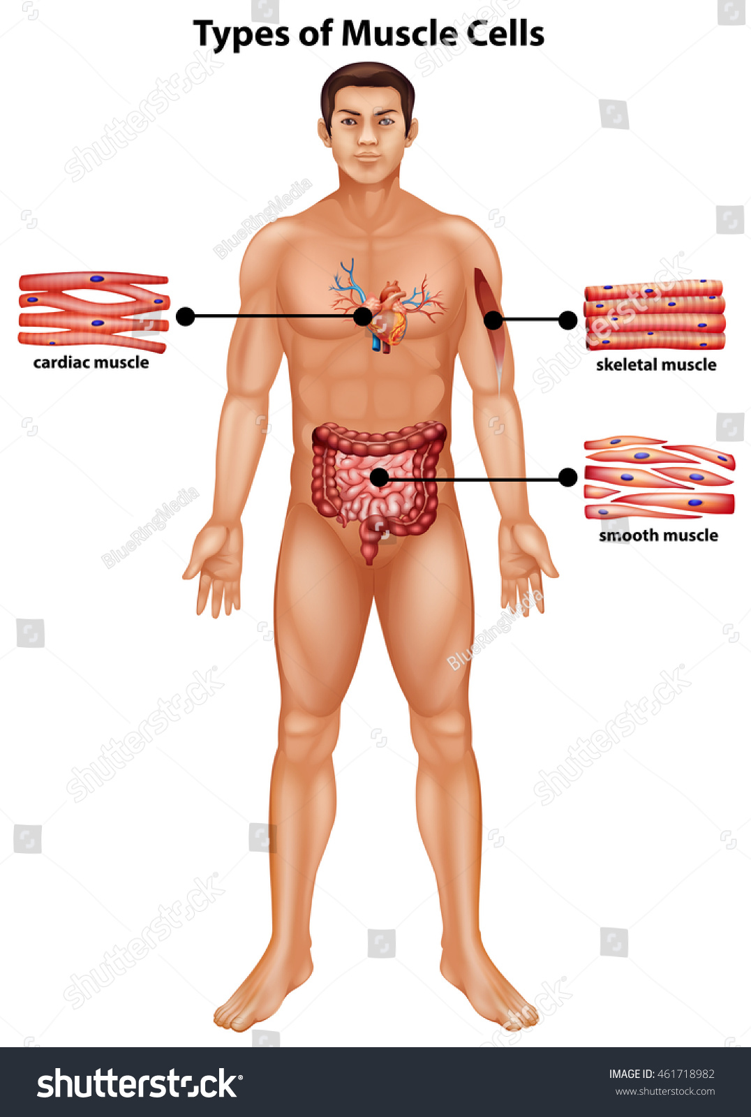
 Diagram showing the types of muscle cells based on location

Functions of the muscle system

some of the key functions of the muscle system:

  1. Movement: Muscles work together with bones, joints, and the nervous system to allow for movement of the body and its parts. The skeletal muscle system is primarily responsible for voluntary movements, such as walking and running, while the cardiac and smooth muscle systems work involuntarily to control the heart and internal organs.
  2. Posture and Stability: Muscles work to maintain posture and stability of the body, helping to keep the body upright and balanced.
  3. Heat generation: Muscle activity generates heat, which helps to regulate body temperature.
  4. Protection: Muscles can also protect internal organs, such as the abdominal muscles that protect the digestive organs.
  5. Circulation: The cardiac muscle system is responsible for pumping blood throughout the body, while the smooth muscle in blood vessels helps to regulate blood flow and blood pressure.
  6. Adaptation: Muscles can adapt to changes in workload, such as during exercise, and can increase in size and strength to meet demand. However, disuse or injury can lead to muscle atrophy and weakness.

Summary

Among other crucial biological processes, muscle contraction aids in posture, joint stability, and heat production. Muscles must contract to sustain positions like standing and sitting. In the human body, there are three different kinds of muscles: Skeletal muscles: These muscles supply the force for movement by being linked to bones.

The muscles that line the insides of internal organs like the stomach and intestines are known as smooth muscles. Cardiac muscles: The heart is made up of these muscles. Additionally, they contract rhythmically and without intentional effort to pump blood throughout the body.


Frequently Asked Questions

1. Describe the purpose of muscle cells.

Muscle cells, also known as fibers, are long, thin cells that are designed specifically to contract. They have protein filaments in them, which use ATP energy to glide over one another. The length of the muscle fibers is reduced or tension is increased as a result of the sliding filaments, which results in contractions. Most bodily motions, both inside and outside, are the result of muscle contractions.

2. Give definitions of muscular atrophy and hypertrophy.

Muscle hypertrophy is an increase in the size of a muscle. While muscle atrophy is a decrease in the size of a muscle.

3. Name the two bodily systems that collaborate with the muscle system to provide movement.

The two bodily systems that collaborate with the muscle system are the skeletal system and the nervous system.

Animal Nervous System

Introduction

Animals have sophisticated nervous systems. It is a vital system that controls all of our body’s voluntary and involuntary movements.  The nervous system controls all complicated functions, including reading, remembering, feeling emotions, and logical thought.  The brain, spinal cord, and nerve network are all parts of the nervous system.

Together, these organs carry out the complex functions of the nervous system.

Nervous System Classification

The nervous system is classified into 3 types i.e the Central nervous system(CNS), the Autonomic nervous system(ANS), and the Peripheral Nervous System (PNS).

Central nervous system(CNS)

  • The spinal cord and brain together make up the CNS. 
  • The brain is covered by a fluid known as a Cerebrospinal fluid(CSF).
  • Three membranes—the dura mater, arachnoid, and pia mater—cover the brain.
  • Humans are made up of the forebrain, midbrain, and hindbrain.
  • The forebrain is made up of the diencephalon and cerebrum and is further divided into the dorsal thalamus and ventral hypothalamus.
  • The cerebrum is in charge of our motivation, reasoning, imagination, memory, thinking, and consciousness.
  • The midbrain is made up of four circular structures called the cerebral aqueduct and corpora quadrigemina.
  • The hindbrain is made up of the cerebellum, medulla oblongata, and pons.
  • All voluntary motions and body equilibrium are controlled by the cerebellum, whereas the pons regulates breathing and the sleep cycle.
  • The medulla oblongata regulates breathing, circulation, gastric secretions, vomiting, and salivation.
  • The spinal cord is a cylindrical structure that resides in the neural canal of the vertebral column.
  • The spinal cord is the location where all the nerves join and the information is sent to the brain.

This image shows the components of Nervous system.

Autonomic nervous system(ANS)

  • ANS is known as the Autonomic Nervous system.
  • It regulates the body’s internal organs’ involuntary functions.
  • The sympathetic and parasympathetic nervous systems are the two types of ANS.

Peripheral Nervous System (PNS)

  • The PNS is made up of nerves that emerge from the spinal cord and brain.
  • The term “cranial nerves” refers to nerves that are emerging from the brain.
  • Spinal nerves are nerves that arise from the spinal cord.

Parts of the nervous system

Neurons are the structural and functional components of the nervous system. Neurons are typically referred to as nerve cells. It is the longest cell in the human body and sends electrical impulses as messages. 

Following are the parts of a neuron.

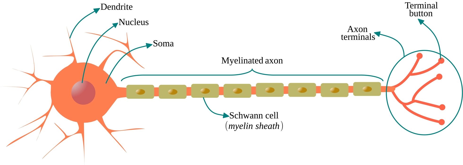
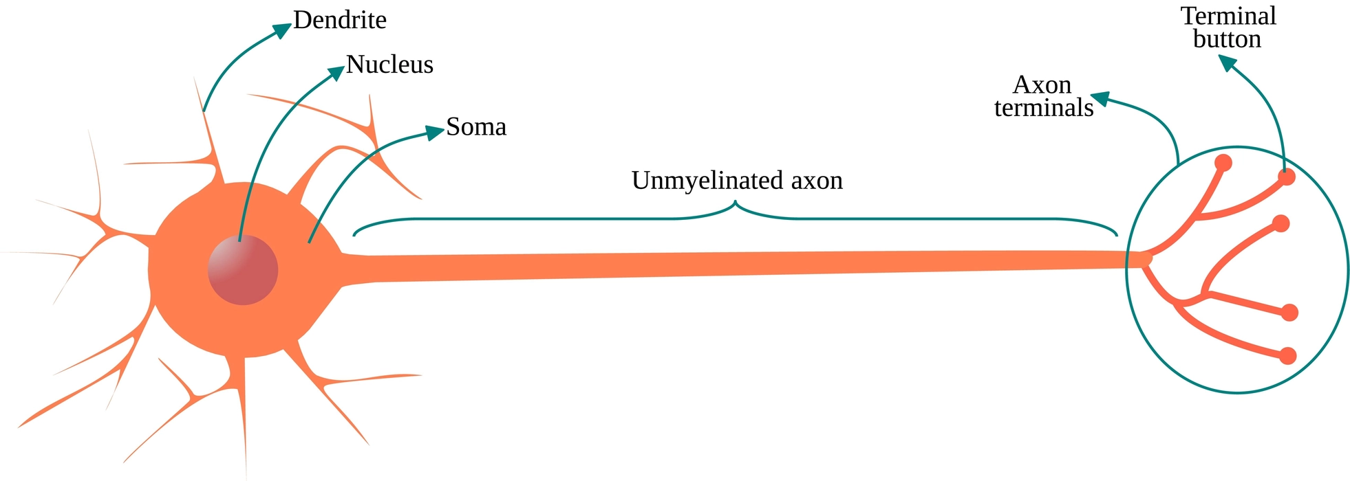
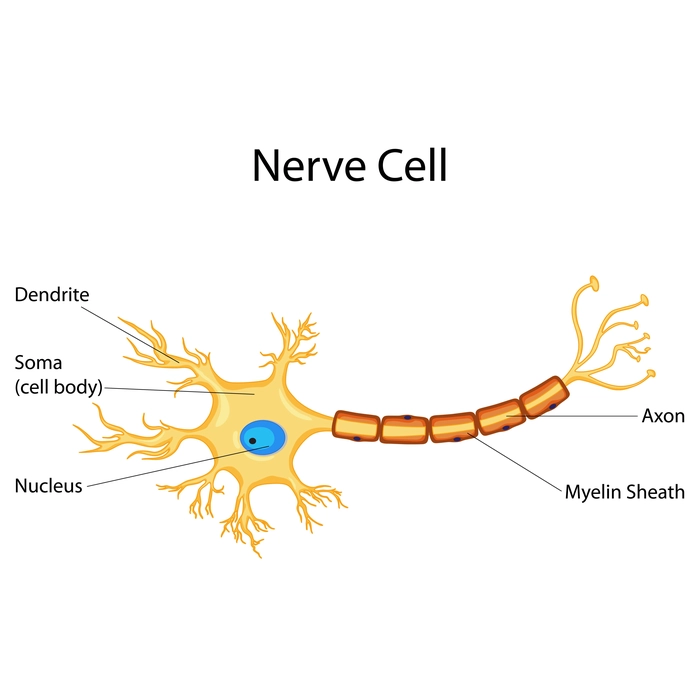
  • Cyton: The cell body is the main component of the neuron. The cyton contains cell organelles, a nucleus, and cytoplasm.
  • Dendrites: These cytoplasmic extensions that protrude from the cell body are numerous and highly branched. They carry electric impulses both inside and outside of the cell body.
  • Axon: It emerges from the cyton as a solitary, unbranched cylindrical projection. A myelin sheath protects the axon. It transports impulses away from the cyton.

Functions of the nervous system

  • The network of neurons is in charge of message reception and transmission, which in turn controls and coordinates the various bodily parts.
  • The cerebrum is in charge of our intelligence, thinking, consciousness, memory, imagination, reasoning, and willpower.
  • The nervous system also controls voluntary movement and body balance. The cerebellum is in charge of this activity.
  • The spinal cord is involved in the reflex action and helps in quick response during dangerous situations.
  • The Nervous system of the body is responsible for controlling all automatic reactions to peripheral nerve stimulation such as breathing, stomach secretion, vomiting, etc.

Types of nerves

Classification of nerves is based on function, structure, and myelin sheath.

Classification based on function

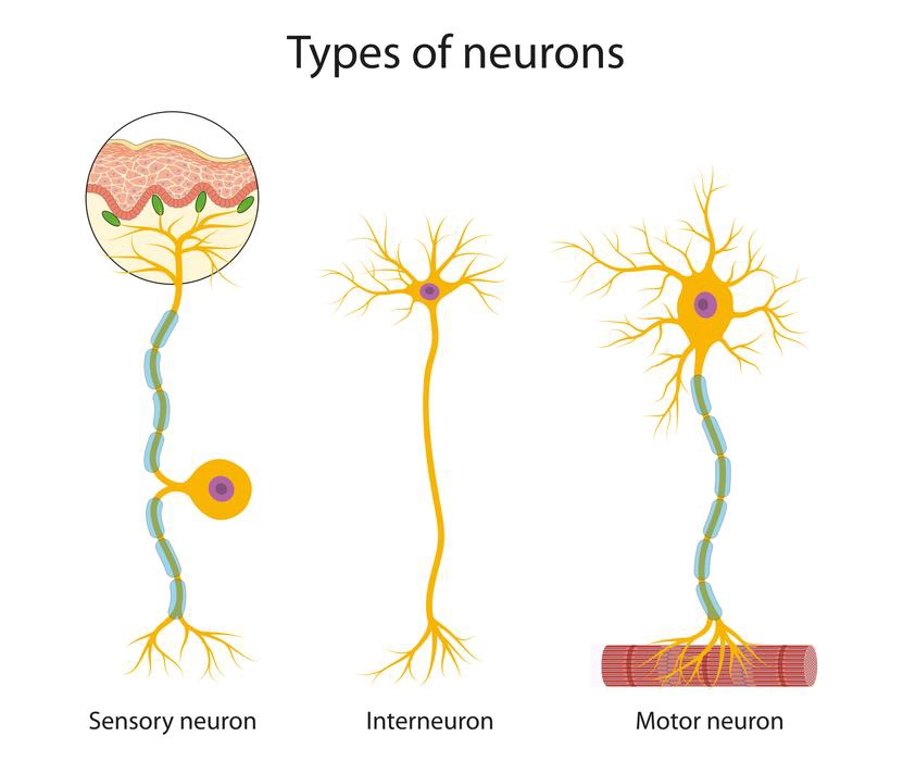
  • Afferent or  Sensory  Neurons: These nerves perform the function of carrying impulses to the Central Nervous system from the various sense organs such as eyes, nose, skin, etc.
  • Efferent or Motor Neurons: These nerves perform the function of carrying impulses from the Central Nervous system to various effector organs such as muscles, organs, etc.
  • Interneurons or Association Neurons: This neuron help in transferring impulses between the Sensory and Motor neurons.

Diagram of types of neurons based on their function.

Classification based on the structure

  • Unipolar Neurons: In these neurons, the cell body gives rise to only a single nerve process that performs the function of both axon and dendron. These are seen in the spinal and cranial nerve ganglia.
  • Bipolar Neurons: Here, two nerve processes are formed from the cell body. One of these becomes the axon and the other becomes the dendron. These are found in various sensory organs such as the nose, tongue, eye, etc.
  • Multipolar Neurons: Multiple dendrons are generated from a single-cell body. These are the most common types of neurons found in the nervous system.

Diagram of types of neurons based on structure.

Classification based on the myelin sheath 

  • Myelinated nerve fiber: A myelin sheath protects the axon 
  • Non-myelinated nerve fiber: The nerve fiber here is not protected by the myelin sheath.

Diagram of structure of myelinated neuron

Diagram of structure of the non-myelinated neuron.

Summary

The human nervous system is the body’s most complicated network system. It is made up of the autonomic, peripheral, and central nervous systems. It enables us to carry out routine tasks, difficult activities, and bodily functions.  With the aid of a sophisticated network of nerves, the nervous system functions as a communication network that sends and receives signals from the brain and spinal cord to every area of the body. The nervous system is made up of neurons and based on their structures, functions, and whether or not myelin sheaths are present, neurons are further classified.

Frequently asked question

1. What is the Cerebrospinal fluid?
Ans: The brain’s ventricles (hollow spaces) are lined with tissue. This tissue produces cerebrospinal fluid. This fluid circulates around and inside the brain and spinal cord to nourish and protect them.

2. What signs of a weak neurological system?
Ans: Symptoms of disorders of the nervous system are-

  • headaches that develop suddenly or persistently.
  • tingling feeling or loss of sensation.
  • loss of muscle strength or weakness.
  • eyesight loss or double vision
  • memory deterioration
  • reduced mental capacity

3. Which components have a negative impact on the nervous system?
Ans: Following have a negative impact on the nervous system 

  • Trauma (injuries), particularly spinal cord and head trauma.
  • Issues that exist from birth (congenital).
  • Mental health issues such as depression, psychosis, or anxiety disorders.
  • Exposure to poisons like lead, arsenic, or carbon monoxide.

Animal Tissues

Introduction

The essential building blocks of living things are cells. There are billions of cells that make up the human body. A group of specialized cells which perform one or more similar functions are referred to as a tissue. They combine to produce several organs, such as the skin, kidney, lungs, liver, and heart. An organ system is made up of two or more organs that perform the same tasks. Each of these contributes to the overall body’s survival.

Types of animal tissues

Animal tissues are classified into four kinds based on their origins, structures, and functions. Animal tissues are of the following types-

  • Muscular tissues
  • Nervous tissues
  • Epithelial tissues
  • Connective tissues

Muscular tissue

Muscular tissue, which is considerably longer and has the ability to contract and expand, which causes motion. These tissues have a lot of blood vessels since they help in activities like running, walking, swimming etc.

Features of Muscular tissues

  • Muscular tissues contract in response to stimuli to produce motion.
  • They can be stretched more than their length.
  • These tissues are elastic in nature because they can expand and then contract to their original length.
  • They can adapt to their surroundings.

Types of Muscular Tissues

This image shows the different types of muscle tissue. They have different characteristics based on the work they do.
TypesLocationFunction
Cardiac muscleHeartContinuous blood pumping
Skeletal muscleSkeletal boneMovement
Smooth muscle Eyes, uterus, blood vessels, and digestive tractMaintain blood flow and blood pressure

Nervous tissues

Nervous tissues make up the central nervous system (CNS) and peripheral nervous system (PNS). By sending nerve impulses, the neural or nervous tissue is in charge of control and coordination of  numerous bodily functions. The spinal cord, the brain, and the nerves are part of nervous system and that stimulate muscle contraction and regulate emotions, memory, and reasoning.

Neurons

Axon, dendrites, and cell bodies make up a neuron, and they are responsible for transmitting and receiving signals throughout the body.

Diagram of nerve cell.

Types of neuronsAccording to their functions, neurons can be divided into the following types:

  • Sensory neurons Short axons and lengthy dendrites are characteristics of unipolar sensory neurons. They are also referred to as afferent neurons since they transport action potential from the sensory receptor to the CNS and brain.
  • Motor neuronsMotor neurons are multipolar and operate as efferent neurons because they transport the action potential from the central nervous system to activate muscles.
  • Associated neurons-Associated neurons help the brain learn, make decisions, and regenerate new neurons. They are multipolar neurons which connect sensory and motor neurons.

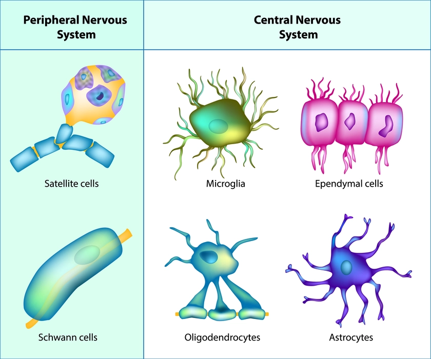
Neuroglial cells 

It is responsible for supporting and maintaining the nervous system and they are of following types-Astrocytes, Ependymal cells, Microglial cells, Oligodendrocytes, Satellite cells and Schwann cells.

This image shows the various types of Neuroglia cells of the nervous system.

Epithelial tissues

All of the body’s inner and outer surfaces are lined and covered with epithelial tissue. The cells form single or several layers and are tightly packed together.

Features of Epithelial Tissues

  • Without intercellular gaps, cells are tightly grouped together to form a sheet.
  • Gap junctions, tight junctions, and adherent’s junctions connect the cells.
  • Despite being non-vascular, or lacking a blood supply, these are innervated (supply nerves to organs or parts of the body).
  • These tissues regenerate rapidly.
  • Although they lack a blood supply, they receive nutrition from substances that diffuse from the blood vessels of the underlying tissues.
  • At the basal surfaces, the epithelial tissues are joined to the connective tissues to forming  basal membranes.
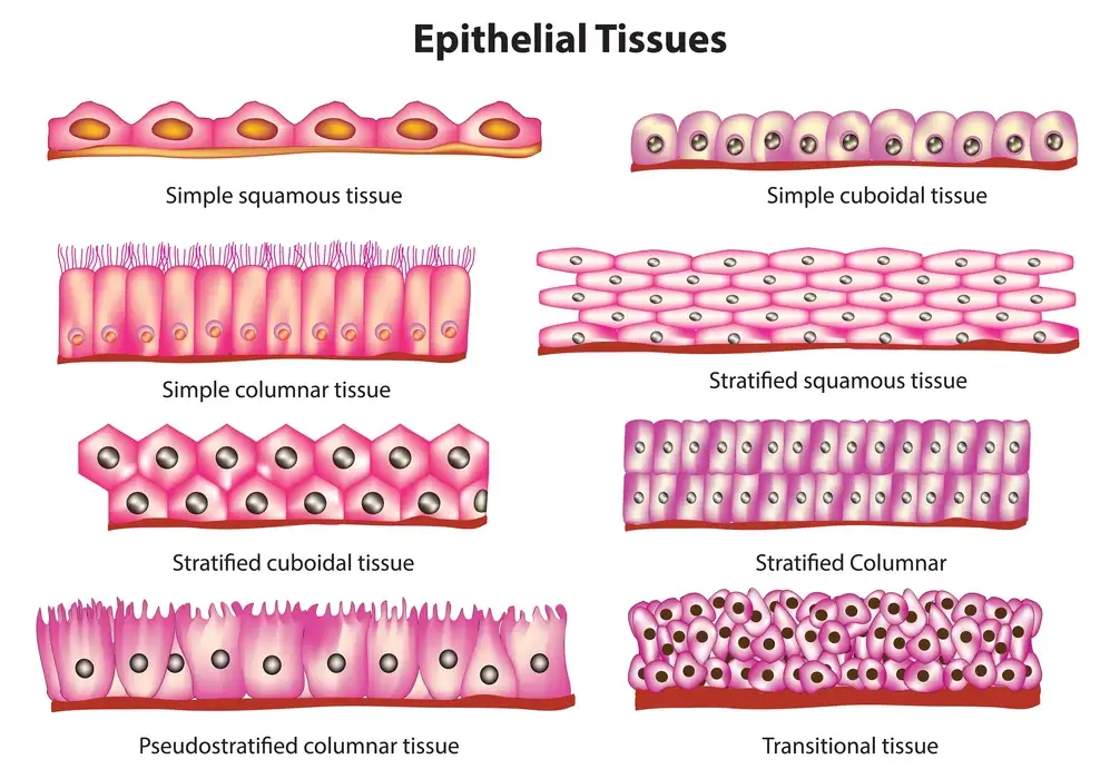
    Types of Epithelial Tissues
TypesLocationFunction
Simple squamousBlood vessels, capillary walls, air sacs, linings of lymphTransport the selective material to pass through osmosis, diffusion, filtration, and absorption
Simple cuboidalLining of ducts, tubular linings of kidneys, surfaces of ovaryAbsorption and secretion
Simple columnarLinings of the respiratory tract, digestive tract and uterusMucous secretion, absorption and protection
Transitional epitheliumInner linings of the ureter, urethra and urinary bladderPrevent reabsorption of toxic materials
Pseudostratified columnar Linings of respiratory passageSecretion, movement of mucous and protection
Stratified squamous Throat, linings of the mouth, vagina, and the outer surface of skinProtection
Stratified cuboidal Salivary glands, mammary glands, pancreas and sweat glandsProtection
Stratified columnar Parts of the pharynx and male urethraSecretion and protection
Epithelial tissues

Connective tissues
Connective tissues connects and supports the body’s all the other tissues and organs. These tissues have the capacity to store fat and aid in the flow of nutrients and other substances between the tissues and organs throughout the body. This transport of nutrients is done thorough the process of diffusion.

Features of Connective tissues

  • Extracellular matrix and cells make up the connective tissue.
  • Mast cells, macrophages, plasma cells, adipocytes, chondrocytes, fibroblasts, osteoblasts, and osteocytes are the cells that present in the connective tissue.
  • Extracellular matrix is made up of tissue fiber and the ground substance.
  • Collagen, elastic, reticular, and fibrillin are the tissue fibers present in the connective tissue. Glycoproteins, glycosaminoglycans, and proteoglycans are ground substances present in the connective tissue.
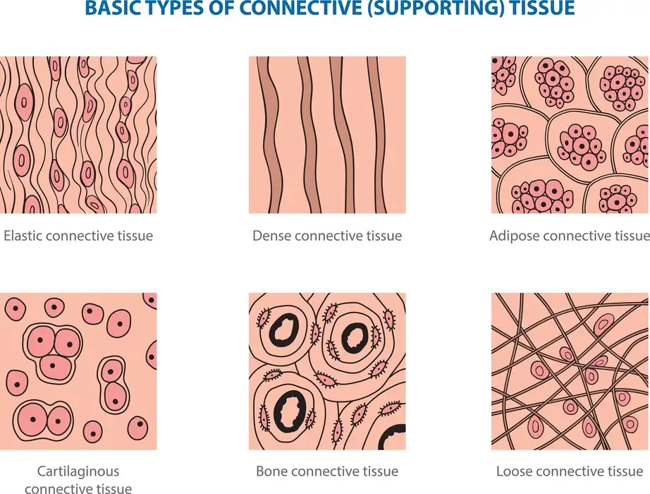
    Types of Connective tissues
TypesLocationFunction
Non-fibrous connective tissues- Blood and adipose tissue.Blood was found throughout the body. Adipose tissue found in association with areolar tissue.Provides nutrition and oxygen to the body. Adipose tissue is a good insulator and source energy reserve.
Collagen fibersTendon, Ligament, Skin, Cartilage, Bone etc.Binding bones and other tissues
Elastic fibersExtracellular matrixGive elasticity to the organs and helps to retain original position post stretching.
Basic types of Connective tissues.

Summary

Animal tissues are categorized according to their shape and function. Tissues are groups of cells having similar functions. Epithelial tissue is present on the upper skin surfaces which have intercellular substances and densely packed cells. Epithelial tissues include squamous, cuboidal, columnar, stratified, transitional, and pseudostratified tissue types. Muscle tissues can contract the muscles to perform particular functions in the heart, skeleton, blood vessels, eyes, and digestive tract. Connective tissues support the placement of organs to enable optimal internal organ form and function. It consists of adipose, areolar, blood, lymph, tendon, ligament, cartilage, bone, and lymphatic tissues. Nervous tissue contains neurons and neuroglial cells that are present  in the brain and spinal cord to control the CNS and PNS and helps entire bodily control and coordination.

Frequently Asked Questions

1. How are various tissues derived?
Ans: All tissues come from one of three distinct germ layers. During embryonic development, these germ layers are formed. They are as follows-

  • The epidermis and nervous system are formed from the ectoderm (outer layer).
  • The middle layer, or mesoderm, eventually forms connective tissue and lines the body’s cavities.
  • The inner layer, or endoderm, gives rise to a number of internal organs, including the pancreas and liver, as well as the lining of the digestive tract, respiratory system, and reproductive system.

2. Give the classification of neurons based on their structure.
Ans: Based on their structural characteristics, neurons can be divided into the following types:

  • Bipolar neurons- Axons and dendrites are two of the extensions seen on bipolar neurons.
  • Unipolar neurons- There is only one extension from the body of a unipolar neuron (one axon).
  • Multipolar neurons- One axon and several dendrites characterize multipolar neurons.

3. What are bones?
Ans: Bones are connective tissue with abundant collagen and calcium, they provide support for the joints that make up the skeletal system of the body. These are places where various lymphocytes of the immune system develop and help in protection of the body.