Acoustic Neuroma

Introduction

Following cell differentiation and proliferation, cell division is the process through which cells are multiplied. Tissues are groups of cells, and the abnormal growth of tissue in an organism is referred to as a tumour. Tumours typically originate as a result of certain disruptions in cell development and the generation of new cells. When a tumour’s growth is restricted, it is benign (non-cancerous), but when it spreads to the body’s key organs, it is malignant (cancerous).

What is Acoustic Neuroma?

Acoustic neuroma is a non-malignant and rare tumour that is also called a Vestibular schwannoma. It is produced by the Schwann cells that surround and support the nerves. The vestibular and auditory nerves, which control balance and hearing, respectively, compose the branches of cranial nerve VIII, commonly known as the vestibulocochlear nerve, where tumours have grown. A critical instance develops when the tumour grows rapidly and continuously.

Causes of Acoustic Neuromas

  • Some people have a rare genetic condition called neurofibromatosis type 2, which is characterized by the formation of tumours on the nerves. Acoustic neuroma is a result of this condition. 
  • Acoustic neuromas are reported in only 5% of patients with neurofibromatosis type 2 (NF2 patients). 
  • In the majority of cases, the exact aetiology of auditory neuroma is unknown. However, some risk variables, including family history, radiation exposure, age, and loud noise exposure, are still thought to be the root cause.

Symptoms of Acoustic Neuroma

Along with other difficulties, the growth of tumours in the vestibulocochlear nerve might affect balance. The following are the symptoms of such tumorous growth:

  • Impaired hearing: Acoustic neuromas 90% of the time accompany some degree of hearing loss. The tumour’s pressure on the nerve or the discharge of compounds harmful to hearing can both cause hearing loss. 
  • Tinnitus: Patients with tinnitus experience a high-pitched hissing or buzzing sound in their ears. Tinnitus can occasionally become persistent. Hearing loss may or may not be present in tinnitus patients.
  • Vertigo and loss of balance: Vertigo, a sudden sensation of the head tilting and spinning, is caused by the growth of a tumour on the balance and auditory nerve. Because of this patient can become unsteady and lurch. 
  • The fullness of the ear: Acoustic neuroma patients may experience full ears as if water is trapped in the ear canal. Hearing loss is frequently to blame for this.
  • Other signs and symptoms of an acoustic neuroma include facial numbness, headaches, nausea, changes in taste, and difficulty swallowing.

Diagnosis of Acoustic Neuroma

The examination of the ear is typically the first step in the diagnosis of an acoustic neuroma, which is then followed by evaluations of the patient’s medical history, imaging, and hearing capacity. Tumours in the brain may be detected with MRI or CT scans using magnetic resonance imaging (MRI) or computerized tomography (CT). The following tests are crucial for determining the presence of an acoustic neuroma: 

  • Audiometry: An audiometer uses a painless hearing test to quantify one’s hearing depending on how loud sounds are and how quickly they vibrate. 
  • Pure Tone Average (PTA): it is a measurement used to assess hearing impairment for speech comprehension. A higher rating denotes a hearing impairment.
  • Speech Reception Threshold (SPT):  The patient can hear speech at this volume at least 50% of the time. A higher score, similar to PTA, denotes hearing impairment.
  • Discrimination in speech (SD): It is a test of the patient’s capacity to distinguish between speech in quiet and noisy settings. Hearing loss is indicated by the lower score.

Treatment for Acoustic Neuroma

The course of treatment for an acoustic neuroma might vary; it is typically determined by the patient’s general health, the size and progression of the tumour, and its symptoms. Three treatment methods are available:

  • Monitoring: Adults who have primary slow-growing tumours may not exhibit any symptoms, making patient surveillance a valuable alternative for follow-up care. The ideal situation for a monitor is when the tumours are up to 1.5 cm in size. Before the tumour grows to a dangerous size, surgery must be performed to remove it.
  • Surgery: Acoustic neuromas can potentially be treated surgically. The surgical procedure’s main goals are to eliminate the tumour and avoid facial paralysis. Complete excision, however, may not always be possible due to the tumour’s proximity to vital brain regions. This procedure carries the potential for several side effects, including hearing loss, tinnitus, cerebrospinal fluid leakage via the nasal route, face numbness, etc.
  • Radiation therapy: It is a non-surgical option; stereotactic radiosurgery, which is most frequently used, can stop the growth of the tumour and lessen the death of neighbouring cells. With this technique, the gamma rays are directed precisely to the tumour without damaging nearby cells. For patients with big tumours, this treatment is not advised.

Summary

Tumours typically originate as a result of certain disruptions in cell development and the generation of new cells. Acoustic neuroma is a non-malignant and rare tumour that is also called a schwannoma. Acoustic neuromas are reported in only 5% of patients with neurofibromatosis type 2 (NF2 patients). Along with other difficulties, the growth of tumours in the vestibulocochlear nerve might affect balance. Tumours in the brain may be detected with MRI or CT scans using magnetic resonance imaging (MRI) or computerized tomography (CT).

 Frequently Asked Questions

1. How does Stereotactic Radiosurgery Work?
Ans. With the help of a 3D coordinate system, stereotactic surgery may find small targets inside the body and carry out a variety of minimally invasive surgical procedures on them, including biopsy, ablation, lesion, stimulation, injection, implantation, and radiosurgery, etc.

2. Define Audiometry?
Ans. A diagnostic hearing test is called audiometry. The loudness of the tone and the speed of the sound determine one’s capacity to hear it. For the detection of hearing impairment, it is crucial.

3. What is the Speech Reception Threshold?
Ans. The speech reception threshold is the lowest degree of speech hearing at which a person can recognize 50% of spoken words. Each ear has reached its speech reception threshold. It serves as a reference point for supra-threshold tests and serves to validate the thresholds discovered using PTA.

4. What is Tinnitus?
Ans. Patients with tinnitus experience a high-pitched hissing or buzzing sound in their ears. Tinnitus can occasionally become persistent. Hearing loss may or may not be present in tinnitus patients.

Adrenal Gland

Introduction

A system of ductless glands that secrete hormones is known as an endocrine gland system. In the blood, hormones are carried directly to their target organs as chemical messengers. As hormones are synthesized in certain tissues, they trigger a cascade of events that allow for certain cellular responses. Adrenal glands are located above the kidney. They are divided into regions cortex and medulla. Both regions produce hormones that regulate electrolyte balance.

What are Adrenal Glands?

The body contains two adrenal glands. These are located above each kidney. They are a part of our endocrine system. Despite their small size, the adrenal glands are quite important for your body’s hormone-related functions. As a result, elements that affect your adrenal glands may have a big impact on your general health. If you believe you might have an adrenal problem, see a doctor.

Want to get an “A” on your Science exams? Let our expert teachers be your guide towards improving your grades and reaching your highest potential. Watch the related video of this chapter in Class 8 Science lesson no 10.

Location of Gland

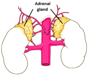
The adrenal glands are endocrine glands that are located over the lateral surface of each kidney’s upper poles. The adrenal glands are located at the back of the abdomen, between the kidney and the ribs. They are triangular, and the parietal peritoneum only covers the front of them. The right gland is pyramid-shaped, while the left gland is moderately-lunar.

Location of adrenal gland


Hormones Secreted by Adrenal Gland 

  • The gland’s outer layer is called the cortex. It generates both cortisol and aldosterone. The medulla is the gland’s most interior area. It generates both adrenaline and noradrenaline.
  • Epinephrine, also referred to as adrenaline, is a hormone that responds swiftly to anxiety by quickening the heartbeat and raising blood sugar levels in the body. 
  • Corticosterone controls the immune system and reduces inflammation in conjunction with hydrocortisone. 
  • Cortisol: With the help of cortisol, the body controls and utilizes fats, protein, and carbohydrates. In addition, it weakens the immune system and affects physiology, which the body uses to help it cope with worry. 
  • Aldosterone: helps the kidneys keep the blood and tissues of the body’s healthy salt chloride level. 
  • Norepinephrine, also referred to as noradrenaline, works in conjunction with epinephrine to respond to anxiety. Its primary function is to prepare the body and mind for combat.

Function

  • The adrenal glands are responsible for producing several hormones that help control blood pressure, produce several reproductive hormones, and maintain the proper level of salt in our blood and cells. 
  • The adrenal glands’ release of stress-inducing hormones causes one of the most well-known reactions, the “conflict of the panic response.” 
  • When and how rapidly an adolescent develops their reproductive system, helping during birth 
  • Our endocrine system’s pituitary gland, also known as the “master gland,” controls our adrenal glands. The main controller of your endocrine glands is the pituitary gland, which is located inside your skull. Abnormal signals may interfere with the number of hormones your pituitary gland advises your adrenal glands to generate.

Disease-related to the Adrenal Gland

Adrenal gland dysfunction might manifest when 

  • When your master gland is unable to efficiently regulate your hormone production, the adrenal glands may form benign or non-malignant tumours or harmful or cancerous tumours. 
  • Can suffer from disorders of the adrenal glands. 
  • People are born with particular gene diseases.

Cushing’s Disease

People with Cushing’s syndrome have excessive cortisol production from their adrenal glands. The most common cause of this is prolonged use of high dosages of corticosteroids. Lupus, arthritis, and pneumonia are just a few of the disorders that are treated with corticosteroids. They have cortisol-like effects throughout the body. 

Symptoms:

  • The round face, the fat accumulation at the base of the neck, and the fat bulge between the arms.
  • Skinny arms and thighs
  • Gaining weight unnecessarily
  • Prone to scarring skin
  • On the hips, chest, and stomach, there are wide, violet stretch marks.

Addison Disease

A rare autoimmune ailment called Addison’s disease may develop when your adrenal glands don’t produce enough cortisol or aldosterone. This self-harm sickness may cause your immune cells to attack the tissues of your adrenal glands. 

Symptoms: 

  • Arterial pressure is high despite the weight 
  • Loss or gain without cause. 
  • Hyperglycemia or intolerance to glucose 
  • low amounts of potassium 
  • Discomfort, stress, or panic episodes

Congenital Adrenal Hyperplasia (CAH)

Extremely little cortisol is synthesized in congenital adrenal hyperplasia (CAH), a genetic disease. Furthermore, persons who have this condition may also have additional hormonal issues in which their bodies create too much testosterone but not enough aldosterone.

Symptoms: 

  • Early acne, body hair, beards, or loud voices (women)
  • Rapid growth in children
  • Sterility due to underarm hair

Adrenocortical Carcinoma

Adrenocortical carcinoma is a malignant tumour that often develops on the exterior of the adrenal gland. Oftentimes, this type of tumour is only found after it has spread to the body’s other systems over generations.

Summary

The adrenal glands are located above the kidney. Despite their small size, the adrenal glands are quite important for your body’s hormone-related functions. The gland’s outer layer is called the cortex. It generates both cortisol and aldosterone. The adrenal glands’ release of stress-inducing hormones causes one of the most well-known reactions, the “conflict of the panic response.” When your master gland is unable to efficiently regulate your hormone production, the adrenal glands may form benign or non-malignant tumours or harmful or cancerous tumours.

Frequently Asked Questions

1. Is it necessary to have both Adrenal Glands to Survive?
Ans. Since the adrenal glands are necessary for human survival, removing both of them (which is quite unusual) forces the patient to take medications and hormone replacements.

2. Is it Possible to live with just one Adrenal Gland?
Ans. Your doctor will administer painkillers to you. Your body can function regularly if your adrenal gland is working properly. If both of your adrenal glands were lost or if your remaining adrenal gland isn’t functioning properly, you might take medication every day to restore the hormones they were making.

3. Do the Adrenal Glands play a role in Digestion?
Ans. The adrenal glands release the stress hormone cortisone to inhibit bodily processes like the immune response and digestion that are not necessary for immediate survival.

4. What are Hormones that make us Drained?
Ans. The adrenal glands also create the hormone aldosterone, which is problematic since it helps regulate your body’s normal fluid and electrolyte balances. As adrenal depletion increases, it makes your body generate less aldosterone, which results in dryness and electrolyte imbalances.