Introduction
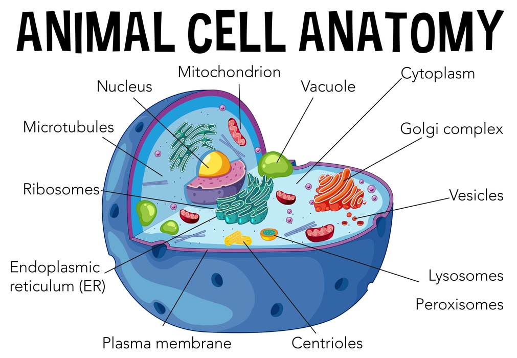
Any living organism’s fundamental structural and functional unit is its cell. In any living organism, cells serve a variety of important tasks in terms of growth, development, and daily activities. They perform these activities through chemical processes that take place inside specialized structures called cell organelles. Eukaryotic cells without a cell wall are referred to as animal cells. Since these cells are eukaryotic, they exhibit the existence of membrane-bound organelles as well as a well-defined nucleus that is protected by a nuclear membrane. It has been noted that animal cells are smaller than plant cells. According to their role in the animal body, they also exhibit a wide variety of sizes and shapes.

Learn More about Animal Cells. Check out more videos in Science Class 8, Lesson 8 – Cell-the unit of life.
Animal Cell Organelles
Cell Membrane
- It is a unique structure that encircles the animal cell and gives it structural stability.
- It controls the movement of various molecules in and out of the cell.
- The characteristic property of the cell membrane is selective permeability.
- A phospholipid bilayer, as well as many kinds of specialized proteins, lipids, and carbohydrates, make up its structure.
- The cell membrane is termed “amphipathic” because the phospholipids of the cell membrane have a hydrophilic head and a hydrophobic tail.
- The cell membrane also plays a critical role in shielding the cell from the outside environment.
- Specialized proteins termed transport proteins aid in the transit of polar molecules through the cell membrane.
Nuclear Membrane
- It can be described as the nucleus’ outer boundary.
- It is the membrane that confines the nuclear area from the cytoplasm It is a double-membrane structure.
- It protects the genetic material from chemical reactions that take place in the cytoplasm.
- The movement of materials into and out of the nuclear area is controlled by the nuclear membrane.
- It consists of nuclear pores, which are points of entry into the nucleus.
- These nuclear pores assist in the transportation of material and help in regulating their movement.
Nucleus
- It is frequently referred to as the cell’s “control center.”
- It is the area where the cell’s genetic material is kept and is in charge of controlling daily cell functions and multiplication.
- It is made up of the nuclear membrane, the genetic material, chromatin, nucleolus, and nucleoplasm.
- The production of ribosomes takes place in the nucleolus.
- DNA, which makes up chromosomes, contains instructions for cell division and growth.
Centrosome
- Animal cells only have these organelles, which are missing from plant cells.
- During cell division, they serve as the main organizing unit for microtubules.
- They are made up of two centrioles, which are two groups of microtubules that are kept perpendicular to one another.
- The development of spindle fibers, which bind to chromosomes in the metaphase and move them toward the poles in the anaphase, is aided by these cells.
Lysosome (Cell Vesicles)
- It is frequently referred to as the cell’s suicide bag.
- These membrane-bound organelles are crucial for breaking down big molecules and eliminating waste from the cell.
- It has an acidic pH and a wide variety of hydrolytic enzymes that aid in its function in the disintegration of molecules.
- Lysosomes develop as budding from the Golgi body, whereas the endoplasmic reticulum makes the hydrolytic enzymes.
Cytoplasm
- It refers to the thick liquid that envelops cellular volume.
- The cytoplasm, includes the cytosol, various organelles (apart from the nucleus), and other macro- or macromolecules.
- The majority of cellular responses and metabolic processes take place there.
- It serves as a matrix in which the other organelles are suspended.
- The fluid nature helps molecules flow more easily inside cells.
Golgi apparatus
- In the Golgi apparatus, molecules created in the endoplasmic reticulum are packaged, modified, and transported to their final location.
- The molecule that has to be delivered is contained in vesicles made by the Golgi system.
- It has two primary faces: the forming face, also known as the cis face, where vesicles attach to be modified in the Golgi cisternae, and the mature face, also known as the trans face, where the vesicles are released.
- Lysosome synthesis is also carried out by the Golgi apparatus.
Mitochondrion
- These are frequently referred to as the “powerhouse” of the cell since they participate in ATP creation.
- This organelle is referred to as a semi-autonomous organelle because it has its own DNA.
- Within this organelle, cellular respiration occurs to finally produce energy.
- It also plays a role in apoptosis, cell communication, and signaling. Other activities include the storage of calcium ions to maintain a balance of calcium ions in the cell.
Ribosomes
- They are small organelles that are not membrane-bound.
- They are mostly located in the mitochondrial matrix, the nucleus, and the cytoplasm.
- They serve as the main location for the synthesis of protein.
- It typically has 2 subunits. It can be distinguished as the 70s and 80s ribosomes in prokaryotes and eukaryotes, respectively.
Endoplasmic Reticulum (ER)
- ER is regarded as the cell’s biggest single membrane-bounded compartment.
- It is made up of a network of vesicles, tubules, and cisterns.
- Here Protein synthesis and protein modification take place.
- The membrane of ER is responsible for the synthesis of lipids and proteins for all organelles of the cell
- Depending on how they appear under the microscope, they are divided into the rough and smooth endoplasmic reticulum.
Vacuole
- It is a unique organelle that is used to store extra cell materials.
- It is surrounded by a tonoplast membrane.
- Water can occasionally be found in vacuoles, which gives the cell a turgor pressure that helps it keep its form and survive adverse circumstances.
Functions of an Animal Cell
The fundamental tasks of an animal cell are-
- Reproduction to insure the continuation of the generation
- Physical growth of the organism.
- Respiration and metabolism fulfill the energy demands of the body.
Animal Cell types
Skin Cells
- These cells are located on the outside of the body and are frequently very thinly layered.
- These cells act as the body’s initial line of defense against germs and various stresses coming from the outside.
- Melanocytes are an example.
Muscle Cells
- These cells have specialized functions that help in the movement of organisms.
- The skeletal, cardiac, and smooth muscles are some examples.
Nerve Cells
- These long, branched cells, also known as neurons, carry electrical impulses throughout the body, aiding in the coordination and control of the entire body.
- Along with neurons, glial and Schwann cells are also present.
Blood Cells
- Red Blood Cells (RBC) and White Blood Cells are the two primary types of Blood cells.
- RBCs utilizing the pigment hemoglobin, function as a transporter of oxygen throughout the body.
- WBCs also referred to as leukocytes, are the body’s defenders that resist and fight infections.
Fat Cells
These large cells, also known as adipocytes, are responsible for storing fat droplets or lipids when the body has an excess of them.
Summary
- Animal cells are made up of various organelles such as Ribosomes, centrosomes, Endoplasmic Reticulum, Nucleus, Nuclear membrane et,c.
- Animal cell types include- skin cells, Muscle cells, Nerve cells, Blood cells, etc.
- Thus, all the cells and their organelles work in unison to bring about proper control and coordination of the entire body.
Frequently Asked Questions
1. Who developed the plasma membrane model?
Ans: Singer and Nicolson presented the fluid mosaic model of the plasma membrane in 1972. According to this, the cell membrane bilayer is made up of phospholipid molecules and proteins are present on the surface and are embedded in the lipid bilayer.
2. What three types of lysosomes?
Ans: The lysosome’s type:
- Primary lysosomes: These are lysosomes that have just formed.
- Secondary lysosomes: When primary lysosomes and phagosomes combine, secondary lysosomes are produced.
- Autophagosomes: They are formed by the digestion of intracellular organelles by the process of autophagy.
3. What are the functions of smooth ER?
Ans: Smooth ER is a membrane-bound organelle that is devoid of ribosomes. The main function of this organelle is the synthesis of carbohydrates, lipids, and steroids. It also performs the metabolism of foreign compounds such as drugs and toxins.

