Introduction
The muscular system is an organ system in the body that is responsible for generating force and movement. It includes all the muscles in the body, as well as their associated tendons, which connect muscles to bones, and ligaments, which connect bones to other bones. The muscular system works together with other systems in the body, such as the nervous system and skeletal system, to coordinate movement and maintain posture. The muscular system is important for a variety of bodily functions, including movement, posture, and heat generation.
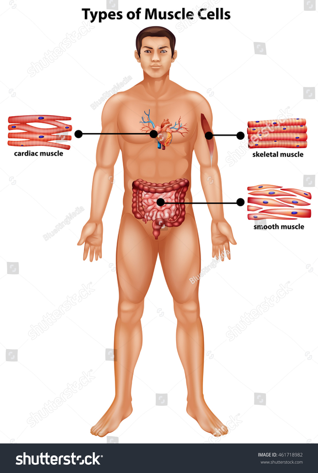
Types of Muscular system
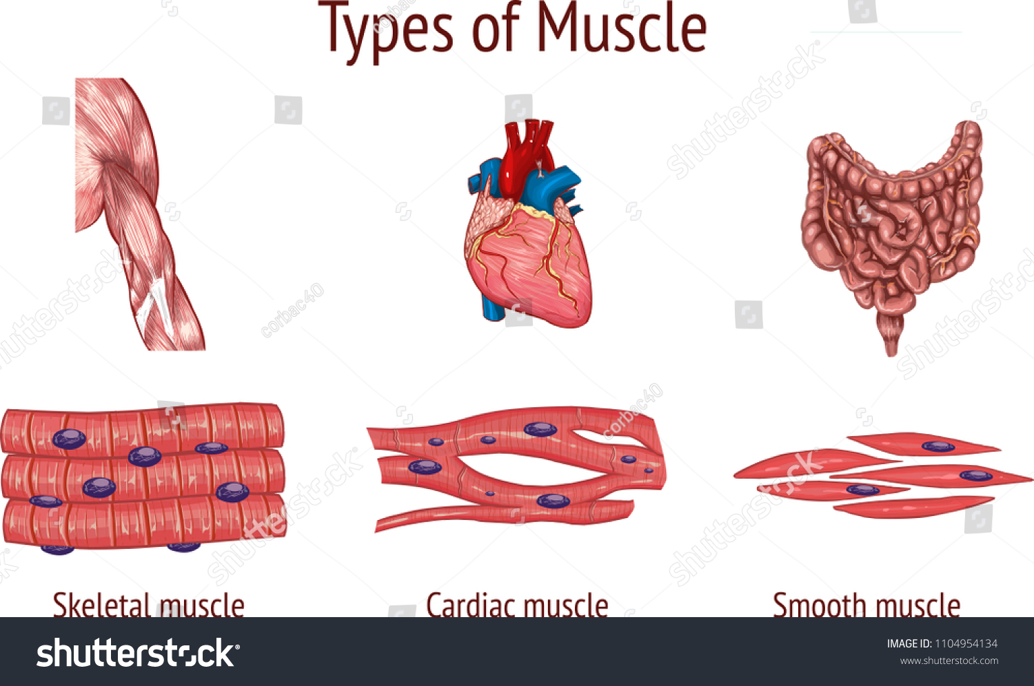
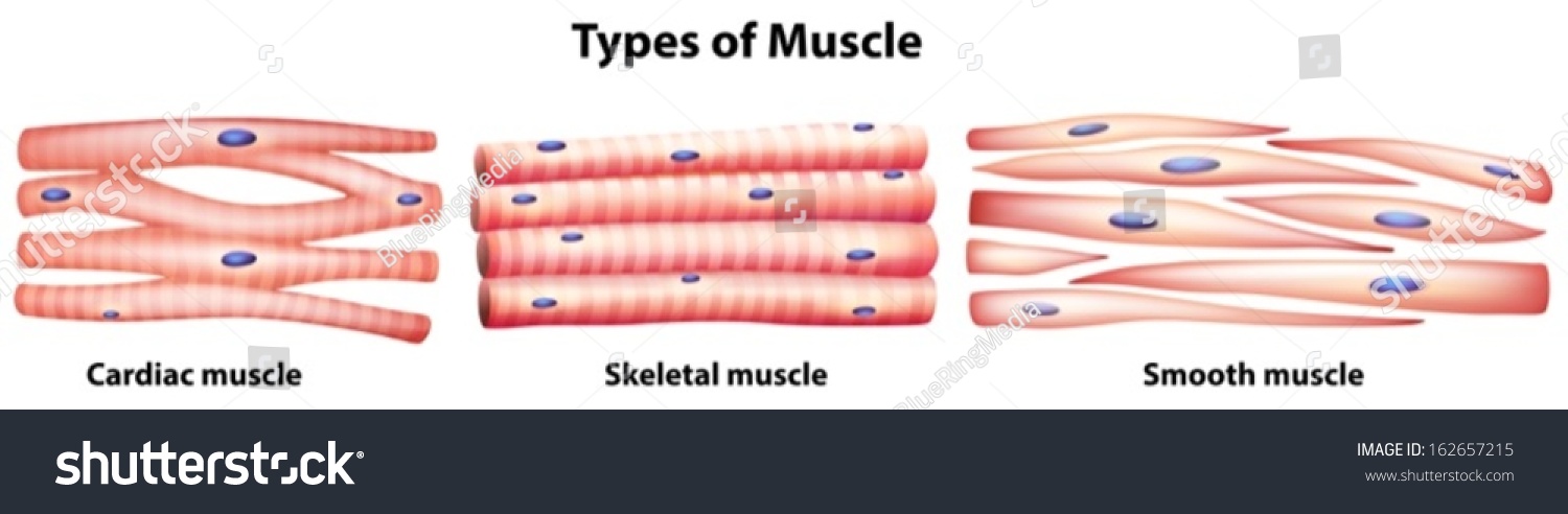
There are three types of muscles in the human body:
- Skeletal muscles
- Smooth muscles
- Cardiac muscles
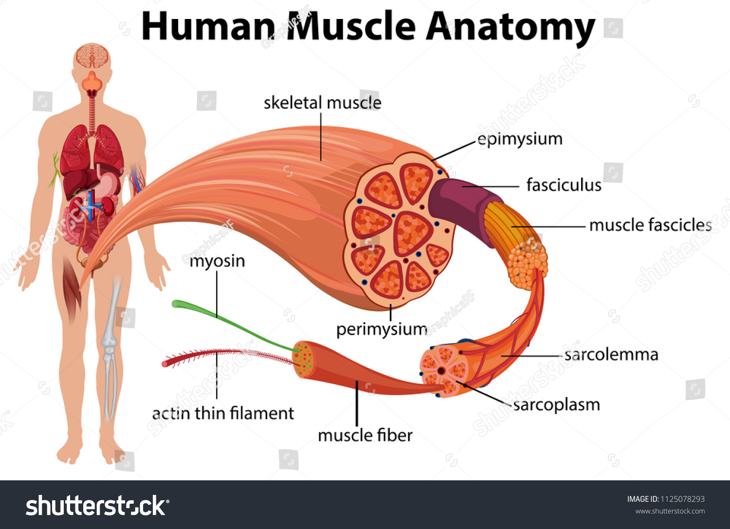
The skeletal muscle system
The skeletal muscle system is responsible for generating movement and providing support for the body. Skeletal muscles are attached to bones by tendons and are under voluntary control, meaning that they can be consciously controlled to move. Here are some key features of the skeletal muscle system:
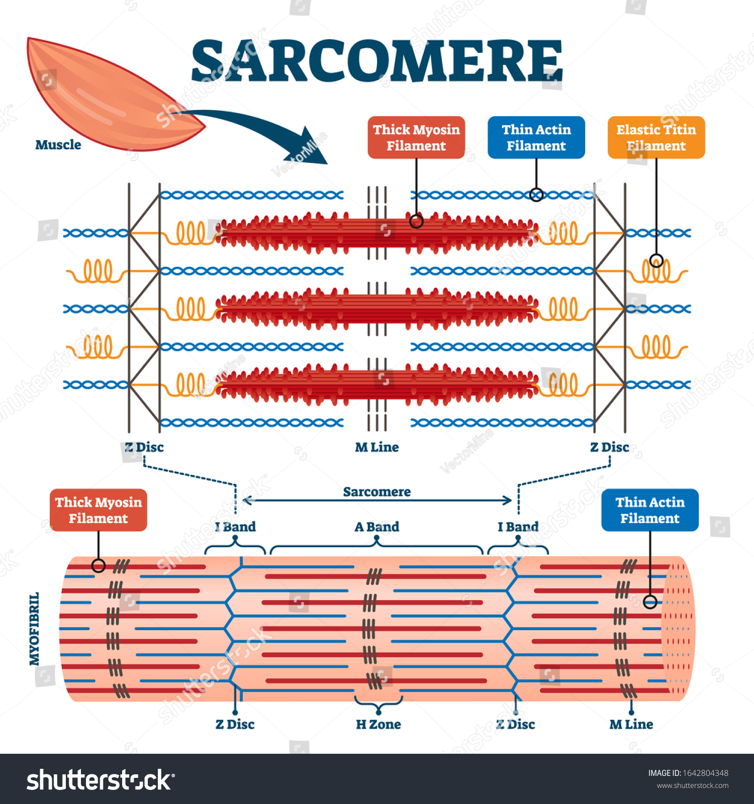
- Structure: Skeletal muscles are composed of bundles of muscle fibers that are surrounded by connective tissue. The muscle fibers are made up of myofibrils, which contain actin and myosin filaments that interact to generate force and movement.
- Function: Skeletal muscles work together with the nervous system to control movement and maintain posture. When a muscle contracts, it generates a force that is transmitted through the tendons to the bones, causing movement. Skeletal muscles can also work in opposition to one another, such as the biceps and triceps muscles in the arm, to produce more complex movements.
- Types of contractions: Skeletal muscles can produce two types of contractions: isotonic and isometric. Isotonic contractions involve movement, such as lifting a weight, while isometric contractions involve no movement but generate tension, such as holding a weight steady.
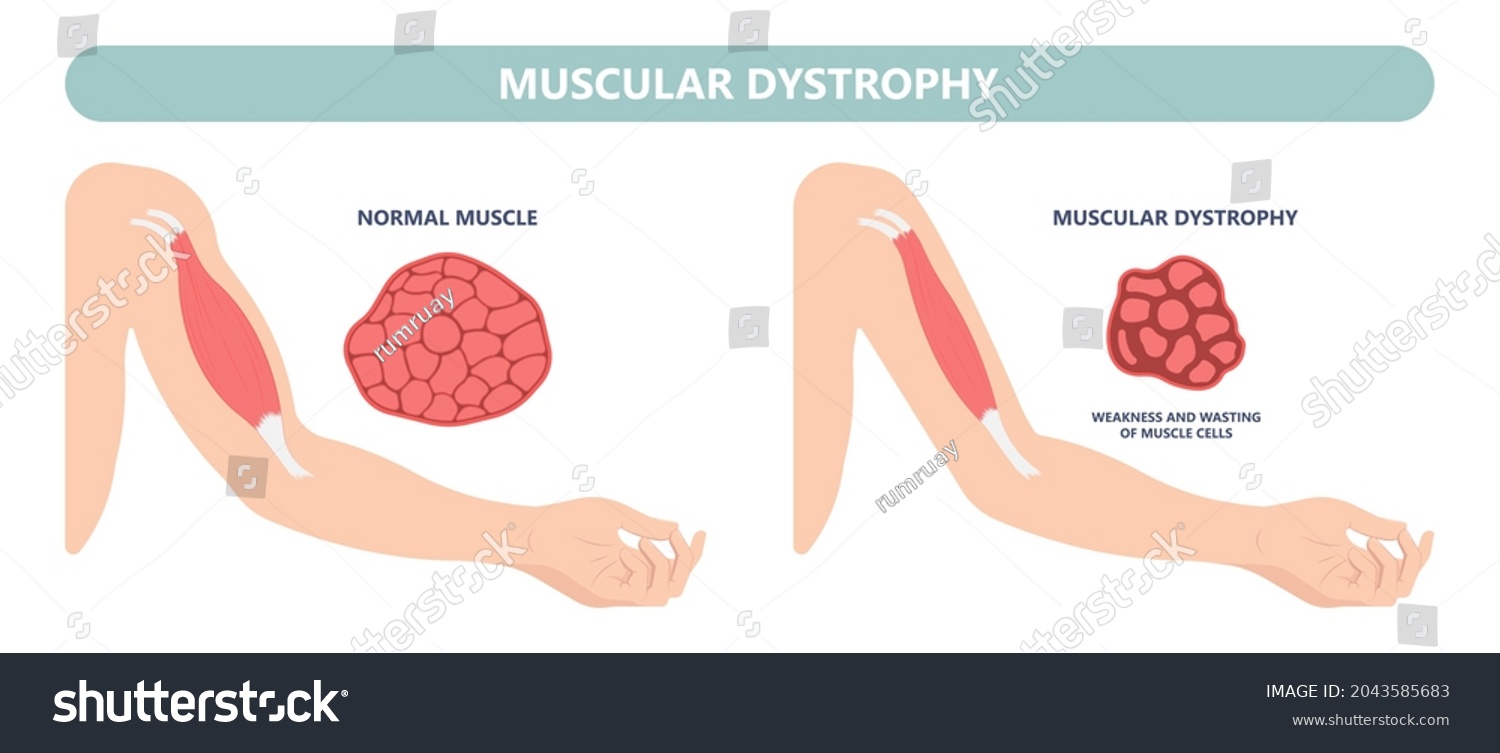
- Adaptation: Skeletal muscles can adapt and change in response to exercise and activity. Regular exercise can increase muscle size and strength, while disuse or injury can lead to muscle atrophy and weakness.
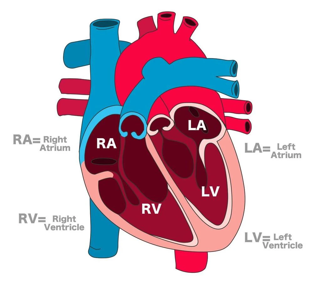
The cardiac muscle system
The cardiac muscle system is the type of muscle that makes up the heart. Unlike skeletal muscles, which are under voluntary control, and smooth muscles, which are not under voluntary control, cardiac muscles are involuntarily controlled and rhythmically contract to pump blood throughout the body. Here are some key features of the cardiac muscle system:
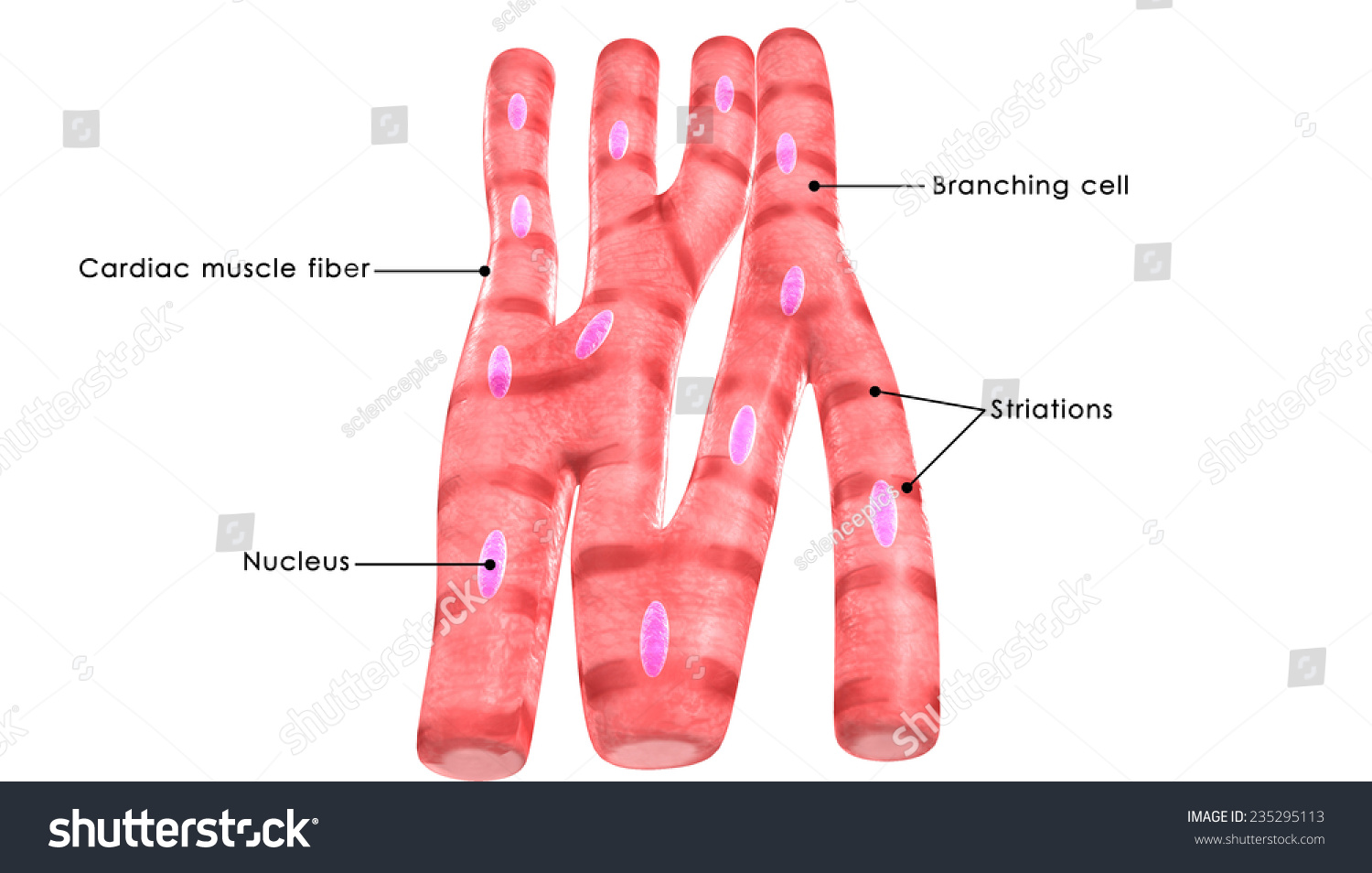
- Structure: Cardiac muscle cells, or cardiomyocytes, are elongated, branched cells that are connected by intercalated discs. These discs contain gap junctions, which allow for the electrical and chemical communication necessary for coordinated contraction of the heart.
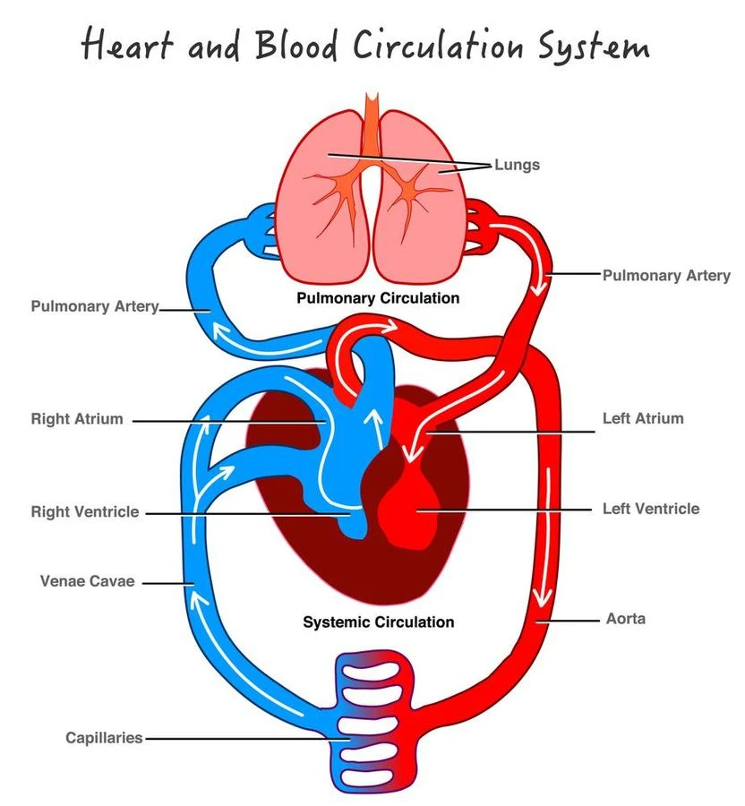
- Function: The main function of the cardiac muscle system is to pump blood throughout the body. The heart has four chambers, and each chamber is lined with cardiac muscle that contracts in a coordinated way to ensure that blood is pumped efficiently.
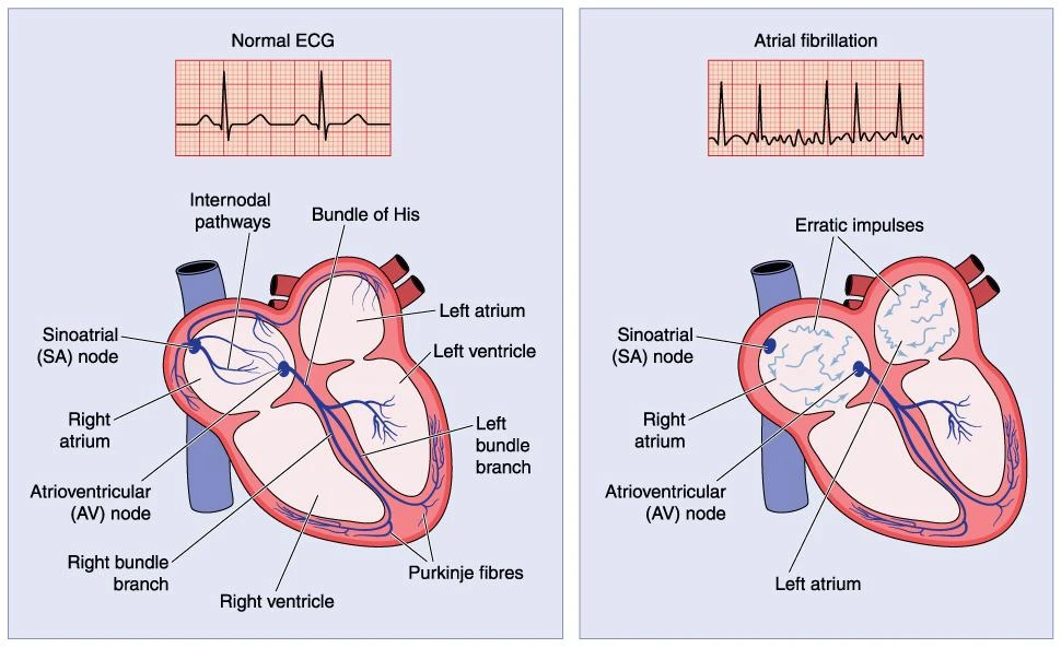
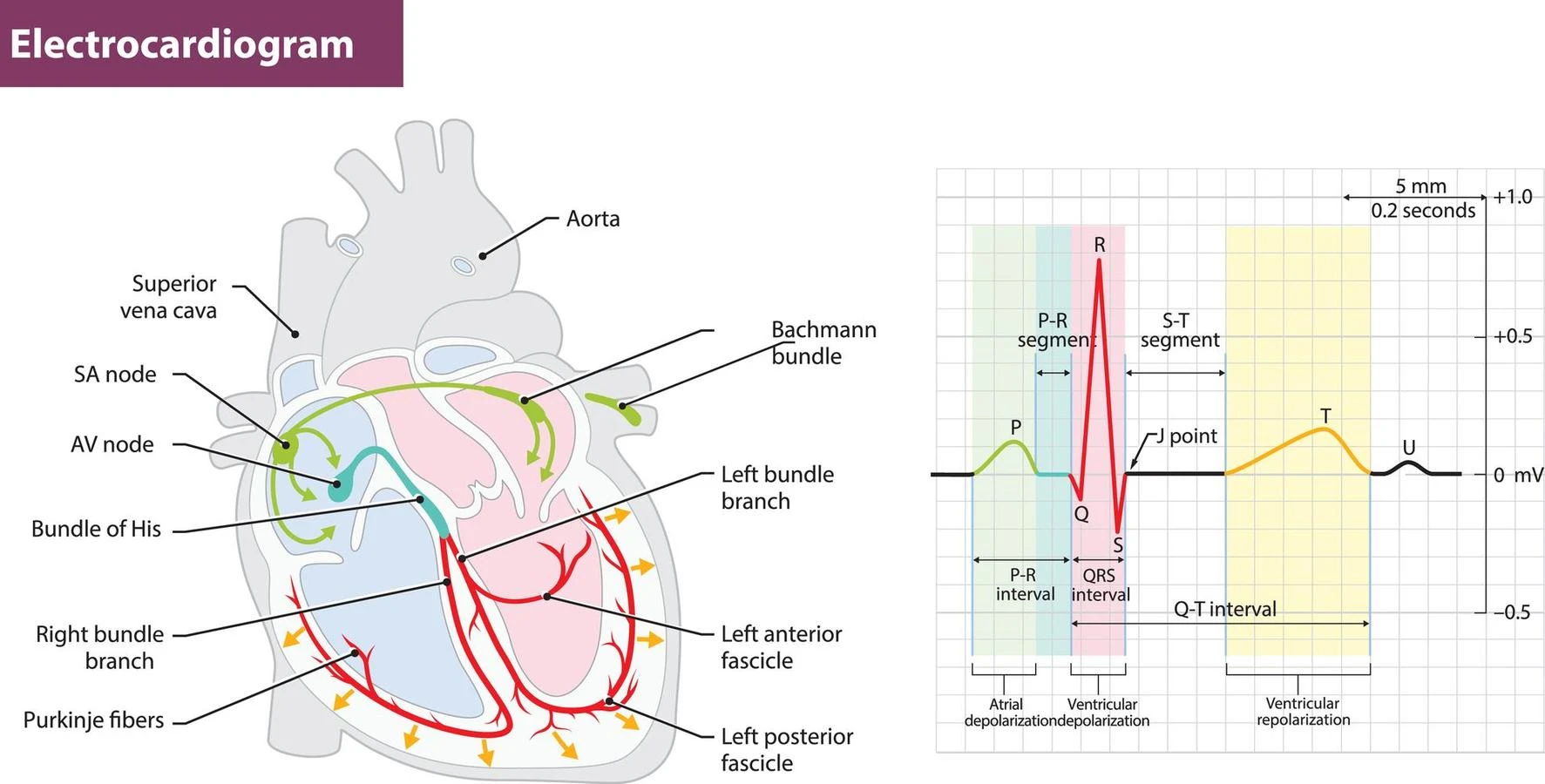
- Electrical control: The electrical signals that control the contraction of the cardiac muscle system originate in the sinoatrial node, which is located in the right atrium of the heart. These signals then spread through the heart’s conduction system, which includes the atrioventricular node and the bundle of His, to ensure that the heart contracts in a coordinated and efficient manner.
- Adaptation: The cardiac muscle system can adapt to changes in workload, such as during exercise or pregnancy, to increase the strength and efficiency of the heart’s contractions.
The visceral muscle system
The visceral muscle system is responsible for the movement of internal organs and structures, such as the digestive tract, blood vessels, and respiratory tract. It is also known as a smooth muscle because of its appearance under the microscope. Here are some key features of the visceral muscle system:
- Structure: Visceral muscle cells, or smooth muscle cells, are elongated and tapered, with a single nucleus. Unlike skeletal muscles, they are not striated, or striped, and do not have the distinct banding pattern of skeletal muscles.
- Function: The main function of the visceral muscle system is to contract and relax to move substances through the body. For example, in the digestive system, visceral muscle contracts to move food through the esophagus and intestines, while in the respiratory system, it contracts to control the diameter of the bronchioles, which affects the flow of air into and out of the lungs.
- Involuntary control: Like the cardiac muscle system, the visceral muscle system is under involuntary control, meaning that it is not directly controlled by conscious thought or action.
- Adaptation: The visceral muscle system can adapt to changes in workload and demand, such as during pregnancy or in response to disease or injury.
Functions of the muscle system
some of the key functions of the muscle system:
- Movement: Muscles work together with bones, joints, and the nervous system to allow for movement of the body and its parts. The skeletal muscle system is primarily responsible for voluntary movements, such as walking and running, while the cardiac and smooth muscle systems work involuntarily to control the heart and internal organs.
- Posture and Stability: Muscles work to maintain posture and stability of the body, helping to keep the body upright and balanced.
- Heat generation: Muscle activity generates heat, which helps to regulate body temperature.
- Protection: Muscles can also protect internal organs, such as the abdominal muscles that protect the digestive organs.
- Circulation: The cardiac muscle system is responsible for pumping blood throughout the body, while the smooth muscle in blood vessels helps to regulate blood flow and blood pressure.
- Adaptation: Muscles can adapt to changes in workload, such as during exercise, and can increase in size and strength to meet demand. However, disuse or injury can lead to muscle atrophy and weakness.
Summary
Among other crucial biological processes, muscle contraction aids in posture, joint stability, and heat production. Muscles must contract to sustain positions like standing and sitting. In the human body, there are three different kinds of muscles: Skeletal muscles: These muscles supply the force for movement by being linked to bones.
The muscles that line the insides of internal organs like the stomach and intestines are known as smooth muscles. Cardiac muscles: The heart is made up of these muscles. Additionally, they contract rhythmically and without intentional effort to pump blood throughout the body.
Frequently Asked Questions
1. Describe the purpose of muscle cells.
Muscle cells, also known as fibers, are long, thin cells that are designed specifically to contract. They have protein filaments in them, which use ATP energy to glide over one another. The length of the muscle fibers is reduced or tension is increased as a result of the sliding filaments, which results in contractions. Most bodily motions, both inside and outside, are the result of muscle contractions.
2. Give definitions of muscular atrophy and hypertrophy.
Muscle hypertrophy is an increase in the size of a muscle. While muscle atrophy is a decrease in the size of a muscle.
3. Name the two bodily systems that collaborate with the muscle system to provide movement.
The two bodily systems that collaborate with the muscle system are the skeletal system and the nervous system.